Author
Rahul Khosla, MD
Assistant Chief, Pulmonary Section
VA Medical Center
Washington, DC
Case
History of present illness: A 40 year old male presents with cough, fever and yellow sputum.
Past medical history: Hypertension
Vital signs: Respiratory rate: 20/min, pulse 85/min, BP 110/80 mmHg, pulse oximetry 96% on room air.
Physical Examination: Decreased breath sounds right lower chest.
EKG: Normal sinus rhythm
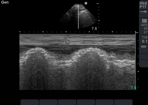
Chest radiograph is pending. In the interim, bedside lung sonography is performed, and the following finding is present on examination of the right lung.
Questions
Question 1: The image is consistent with the presence of which of the following:
A. Pleural effusion
B. Pneumothorax
C. Pneumonia
D. Pulmonary embolism
Question 2: The image can be best described as:
A. Bat sign
B. Sinusoid sign
C. Curtain sign
D. Seashore sign