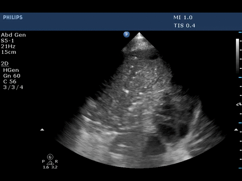
The ultrasound image above shows dynamic air bronchogram.
Portable bedside chest X-ray in the present case showed alveolar
consolidative pattern. Based on X-ray, it is difficult to differentiate
atelectasis from pneumonia. Bedside ultrasound can be a valuable tool
to differentiate between these conditions.
Acute alveolar consolidation is characterized by the tissue-like sign
or shred sign. The tissue-like sign is appearance of lung similar to
liver or spleen, which is echoic with trabeculations and seen in
trans-lobar consolidation. The shred sign appears at the contact point
of consolidated lung with aerated lung as an irregular hyper-echoic line
and seen in non-trans lobar consolidation. The sensitivity and
specificity of ultrasound to detect acute alveolar consolidation, when
both these signs are present is 90%and 98% respectively. The acute
alveolar consolidation can be from pneumonia or resorbtive atelectasis.
Air bronchogram is described as a punctiform or linear hyperechoic
artifacts within the consolidation. It is considered dynamic if there is
>1mm movement with air entry and is due to centrifugal movement of
gas bubbles in bronchus, a sign of airway patency. The specificity and
positive predictive value of dynamic air bronchogram in presence of
alveolar consolidation to predict pneumonia is 94% and 97% respectively.
-
Lichtenstein D, Mezière G, Seitz J. The dynamic air bronchogram. A
lung ultrasound sign of alveolar consolidation ruling out atelectasis.
Chest. 2009 Jun; 135(6):1421-5.
-
Lichtenstein D, Lascols N, Mezie` re G, et al. Ultrasound diagnosis
of alveolar consolidation in the critically ill. Intensive Care Med
2004; 30:276–281.
-
Lichtenstein D, Mezie` re G. Relevance of lung ultrasound in the
diagnosis of acute respiratory failure: the BLUE-protocol. Chest 2008;
134:117–125
-
Lichtenstein D. Lung ultrasound in the critically ill. Ann Intensive Care. 2014 Jan 9;4(1):1.