Authors
Ghassan Kamel, Armin Krvavac, Pujan H Patel, Christopher Barrios, David Stoeckel
Division of Pulmonary, Critical Care and Sleep Medicine, Department of Internal Medicine,
Saint Louis University School of Medicine, Saint Louis, MO
Case
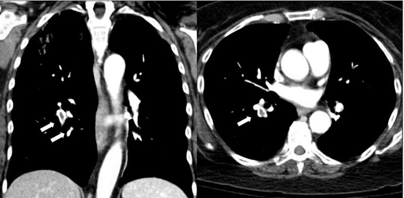
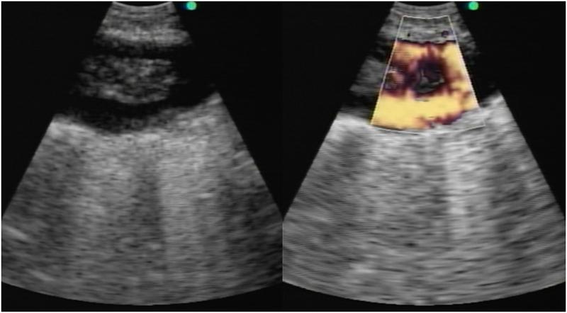
A 57-year-old female with a history of rheumatoid arthritis on chronic prednisone and methotrexate was admitted to the hospital with acute hypoxic respiratory failure. She was initially admitted to the medical intensive care unit (ICU) with a diagnosis of pneumonia but she did not improve with antibiotics. Given her risk factors for opportunistic infections, a diagnostic bronchoscopy with linear endobronchial ultrasound (EBUS) was performed. An abnormality in the pulmonary artery was noted (Figure 1).
Figure 1
Question
What is the abnormality observed in the images above?