Author
Andrew Badke, MD
Pulmonary and Critical Care Medicine Fellow
University of Utah School of Medicine
Case
Presentation
A 59 year-old female was discharged 10 days ago after receiving anticoagulation for a subsegmental pulmonary embolism that was provoked by surgical fixation of a tibial fracture. Over the subsequent weeks she began to develop increasing dyspnea on exertion, nonproductive cough, nausea, and generalized malaise. Prior to her hospitalization, she reported several months of mild dyspnea with exertion and intermittent lower extremity edema. On admission, she presented tachypneic, somnolent, and toxic appearing.
Physical exam
BP: 97/57, HR 97, RR 24, Sa02 86% on room air.
Mild scleral icterus, jugular venous distension, bibasilar crackles, regular rhythm without murmurs; bilateral pitting edema to thighs, cool extremities.
Hospital Data
BUN 68 mg/dL, Cr 1.43 mg/dL, ALT 3269 U/L, AST 4150 U/L, Bilirubin 3.6 mg/dL, INR: 8.2, WBC 13.8 k/µL
troponin 0.07 µg/L, BNP 2,493 pg/ml, lactate 11.2 mmol/L
EKG: Sinus rhythm with a normal axis and a previously documented left bundle branch block
Chest Radiograph: Bilateral pleural effusions, hilar congestion, cardiomegaly
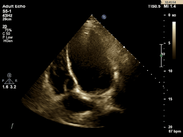
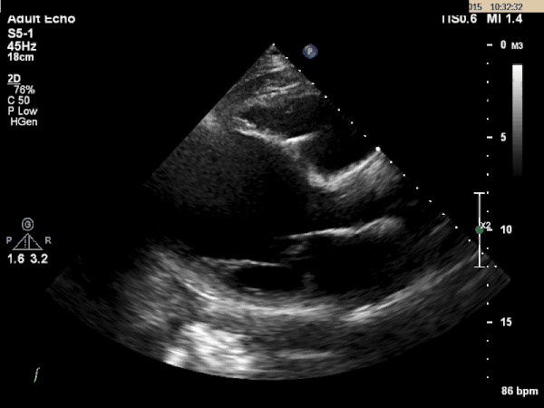
Echocardiogram
Question
What is the most likely etiology behind this patient's multi-organ dysfunction?