Author
R.G. Thomas MD1, M.E. Hurwitz MD1, S.T. Maurer MD2, D.P. Gibson MD3, D.N. Homnick MD, MPH1.
1Pediatric Pulmonology, Michigan State University, East Lansing, MI. 2Pediatric Cardiology, University of Michigan, Ann Arbor, MI. 3Pediatric Radiology, Beaumont Children’s Hospital, Royal Oak, MI.
Case
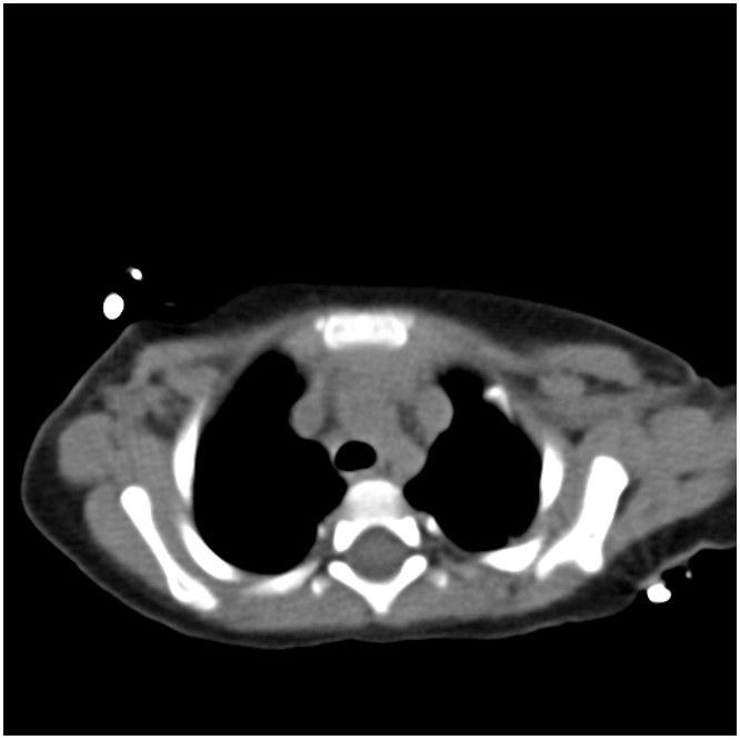
A 12-month-old boy presented for evaluation of recurrent bronchiolitis and hypoxemia. He was repeatedly admitted for acute respiratory failure at age 6, 9 and 11 months old. At 6 and 9 months of age he was positive for RSV but there was no clear infectious source in the third admission. He required oxygen support with all admissions. He had a normal echocardiogram at 2 and 11 months of age. At 13 months old he was seen by his pediatrician and noted to have an oxygen saturation of 88% on room air with no clear inciting illness which prompted another admission and pulmonary consultation. CT of the chest demonstrated the following:
Question
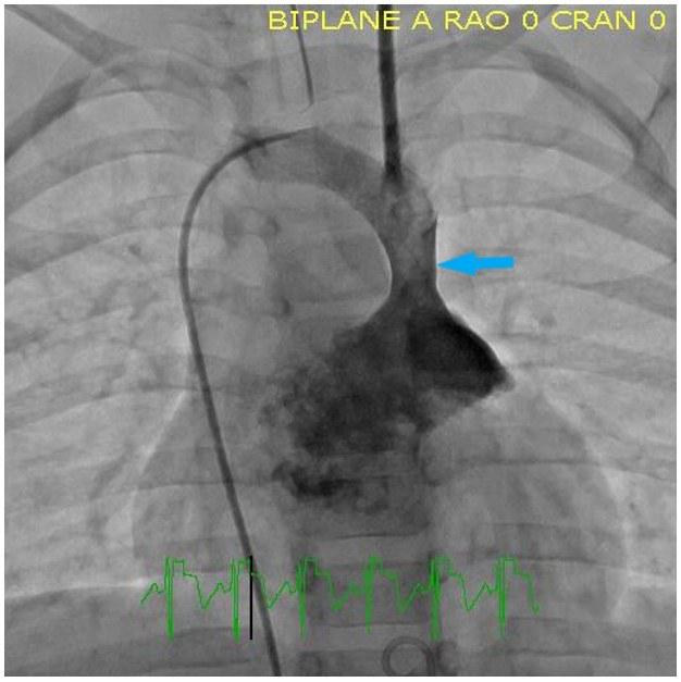
What abnormality does the above chest CT reveal? What is the next best test to evaluate this abnormality?
A. Right sided aortic arch and thoracic MRI
B. Persistent left superior vena cava and cardiac catheterization
C. Mediastinal mass and PET scan
D. Tracheomalacia and bronchoscopy